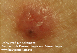
Picture 1: A small nodular type basalioma
The basal cell carcinoma (BCC) is the most frequent type of malignant epithelial tumors of the skin. It is also described as “semi malignant” reflecting on the one hand the capacity to grow invasively even destroying skeletal structures analogous to malignant tumors and on the other hand, in contrast to other malignant tumors, the rare event of mestastasis (seeding into distant organs which is an important characteristic of cancer). Metastasis occurs indeed only in very advanced stages when a BCC has not been treated for a very long time, i.e. years to decades. When a BCC is not treated for many years, they can become noticeably big and are then often covered by the patient with clothes or bandage. Similar to squamous cell carcinomas (SCCs), BCCs are skin colored and surgery in healthy skin is sufficient. Therefore, SCCs and BCCs together are often called “non-melanoma skin cancer”.
For arranging an appointment for treatment of a Basal cell carcinoma in Vienna, please contact our office by phone, email ordination@hautarztokamoto.at or use the online-form.
What is the origin of basaliomas?
As the name indicates, BCCs evolve from cells of the basal layer of the epidermis. They share similar features with squameous cell carcinomas, however, BCCs do also occur on skin that is not consistently exposed to the sun. Although they evolve frequently on sun exposed areas, they can be found in otherwise protected areas as well, such as the trunk.
Features – how do basaliomas look like?
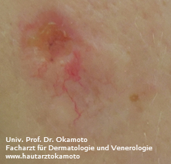
Picture 2: elevated skin colored tumor with widened vessels (teleangiektasia).
Basaliomas share a typical pearlescent surface with widened capillaries (teleangiectasia). They come with various shapes but at a closer look, the lesions consist of nodule(s). Sometimes, they can appear like a red patch resembling an eczema. Often, these nodular parts surround a central ulcer like a wall. These ulcers can bleed, forming a crust which is often scraped off. Patients might report that he or she has scratched that particular spot which has lead then to bleeding, not knowing that in fact the lesion had formed a crust even before the patient started noticing it.
Prevention
Equal to other types of skin cancer, sun is the most important exogenous risk factor of BCCs. Particularly those with a light skin complexion and poor tendency to tan should be more cautious. Particularly patients who have been diagnosed for BCC or any other types of skin cancer (melanoma or SCC) should be more careful and follow up checks in regular intervals are recommended.
Treatment (as of March 2016)
Small and superficial (non-invasive) basaliomas or BCCs are commonly treated with liquid nitrogen (“freezing”). After the treatment, the treated area forms a blister, which heals like any other blister on the skin (such as one on the heal after wearing new shoes). It is an easy way to remove superficial lesions (also used for actinic keratosis and warts).
However, Invasive BCCs should be removed surgically. Only a histologic examination of the tissue warrants the correct diagnosis and the removal of the entire tumor surrounded by healthy skin. Are there multiple smaller BCCs, photodynamic therapy can be considered as an alternative to surgery. The BCCs are treated with a sensitizer before irradiation with UV light. Neoplastic cells that form the tumor are then more sensitive to UV light compared to normal cells and die off. This strategy could safe surgery, however this is not suitable for all patients, and frequent follow up checks are required. Only surgery can provide safety with regard to diagnosis and complete removal.
In case surgery is not possible due to the size or the location of the tumor, a new drug called vismodegib has become available only recently. It is an inhibitor of a specific signaling pathway of the tumor cells and is applied orally. Therefore, no hospitalization is required for therapy. In any case, follow up checks of the entire skin by a specialist are required in regular intervals after treatment.
Common non-cancerous tumors of the skin
Fortunately, most alterations on the skin are benign and do not require treatment. Those are the most common types of benign lesions:
- moles
- seborrhoic keratosis (“age spots”)
- hemangiomas
- lipoma
- warts
Please note
The content of this homepage is intended to help patients to obtain basic information about the above described topics and does not claim to be complete. Diagnosis and therapy is individual and can differ in certain circumstances from the above described. In case of any doubts or further questions, please ask your doctor.
Contact Prof. Dr. Okamoto
For appointments, please contact us by phone, or via E-Mail at ordination@hautarztokamoto.at or contact form.